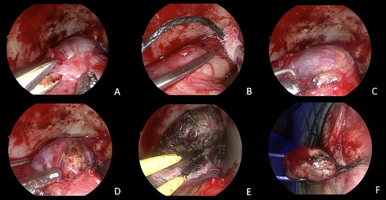
Figure 3.
Intraoperative images. An endoscopic transorbital superior eyelid approach was performed. Sharp (A) and smooth (B,C) dissection of the CVM from the periorbita, until entire exposure of the lesion is achieved (D). Gentle coagulation of the capsule with bipolar forceps (E). En bloc resection of the CVM (F).

