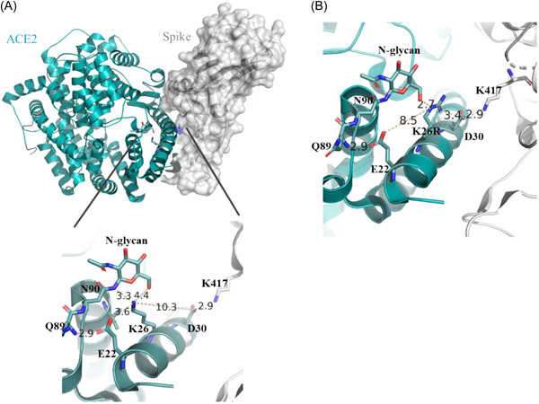
Figure 1.

Structure of ACE2‐Spike (6LZG). (A) ACE2 is shown as a cyan cartoon and Spike is shown on a white surface. At the zoom‐in structure, Lys26 interacting amino acids are shown. (B) In silico generated Lys26Arg ACE2 mutation and its proposed interaction scheme is shown.
