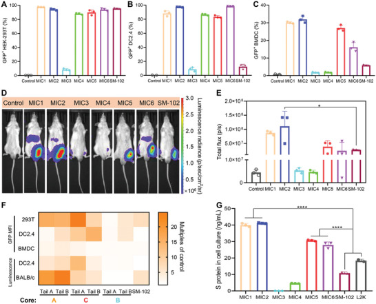
Figure 3.

Expression of the reporter gene and DS mRNA delivered by 4N4T‐LNPs. A–C) 293T cells, DC2.4 and BMDCs were transfected with 4N4T‐GFP mRNA and then harvested after 24 h. GFP+ cells were analyzed and quantified using flow cytometry. D,E) In vivo expression of FLuc mRNA delivered by 4N4T‐LNPs. Male BALB/c mice were intramuscularly injected with 20 µg of FLuc mRNA encapsulated in 4N4T‐LNPs. Bioluminescence was measured 8 h later in an IVIS imaging system. F) MFI of GFP and value of bioluminescence were all converted into multiples of the negative control. Core A: MIC1, MIC2; Core B: MIC 5, MIC6; Core C: MIC3, MIC4. G) The concentration of S protein in the cell culture was measured using ELISA after 293T cells had been incubated with 4N4T‐DS mRNA vaccines for 24 h. All data are presented as the mean ± SD (n = 3). Statistical significance was analyzed by one‐way ANOVA. (ns or unmarked, not significant; *, p < 0.05; **, p < 0.01; ***, p < 0.001; ****, p < 0.0001).
