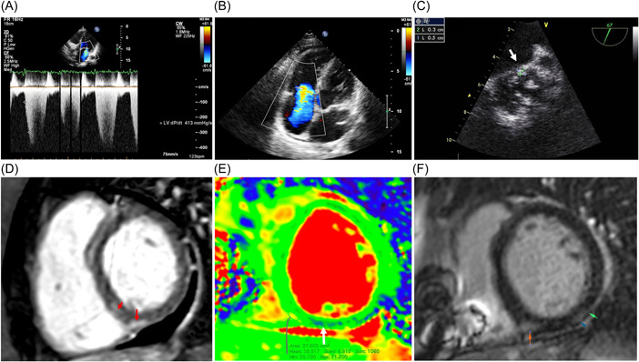
Figure 1.

Echocardiography and magnetic resonance imaging in patients with post‐COVID myocarditis. Upper series—echocardiography: (A) decreased dp/dt (413 mmHg); (B) severe tricuspid regurgitation due to dilatation of the right ventricle; (C) vegetation on the bicuspid aortic valve measuring 3 × 5 mm (arrow), transesophageal study. Lower series—MRI: (D, F) late gadolinium enhancement in the posterior septal and posterior segments of the left ventricle (arrows); (E) edema along the posterior septal segment of the left ventricle (T2 map).
