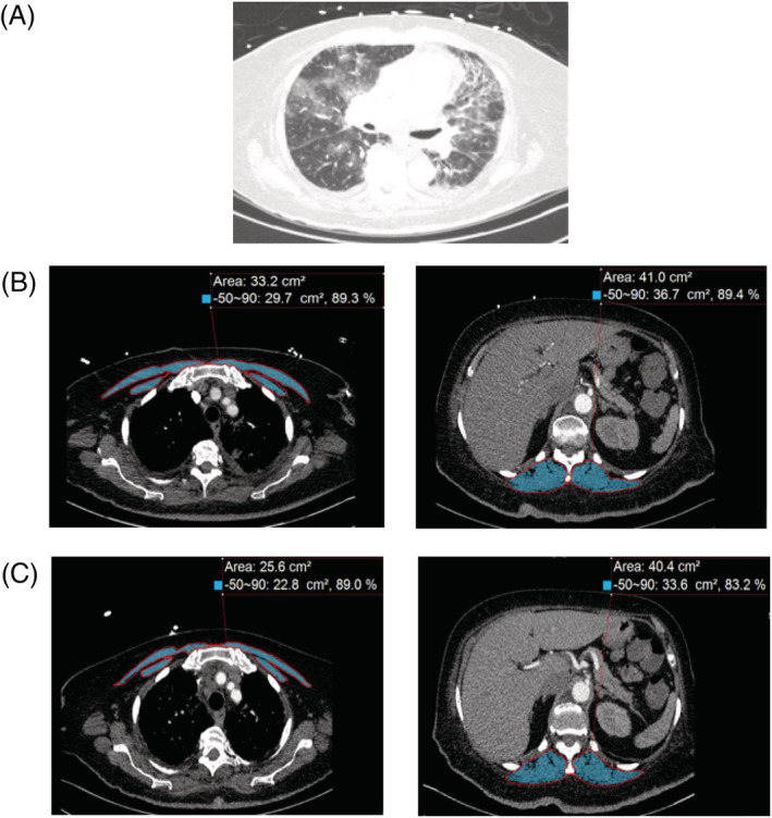
Figure 2.

Representative computed tomography (CT) scans at thoracic level used to determine muscle area in patients with COVID‐19. (A) Representative CT image utilizing lung windows demonstrates evidence of COVID‐19 related bilateral pneumonia. (B) Representative CT image for pectoralis muscle and erector spinae muscle imaging from the initial CT scan are shaded. Skeletal muscle CSAs are measured in cm2. (C) Representative CT image for pectoralis muscle and erector spinae muscles from the subsequent CT scan are shaded. Skeletal muscle mass CSAs are measured in cm2.
