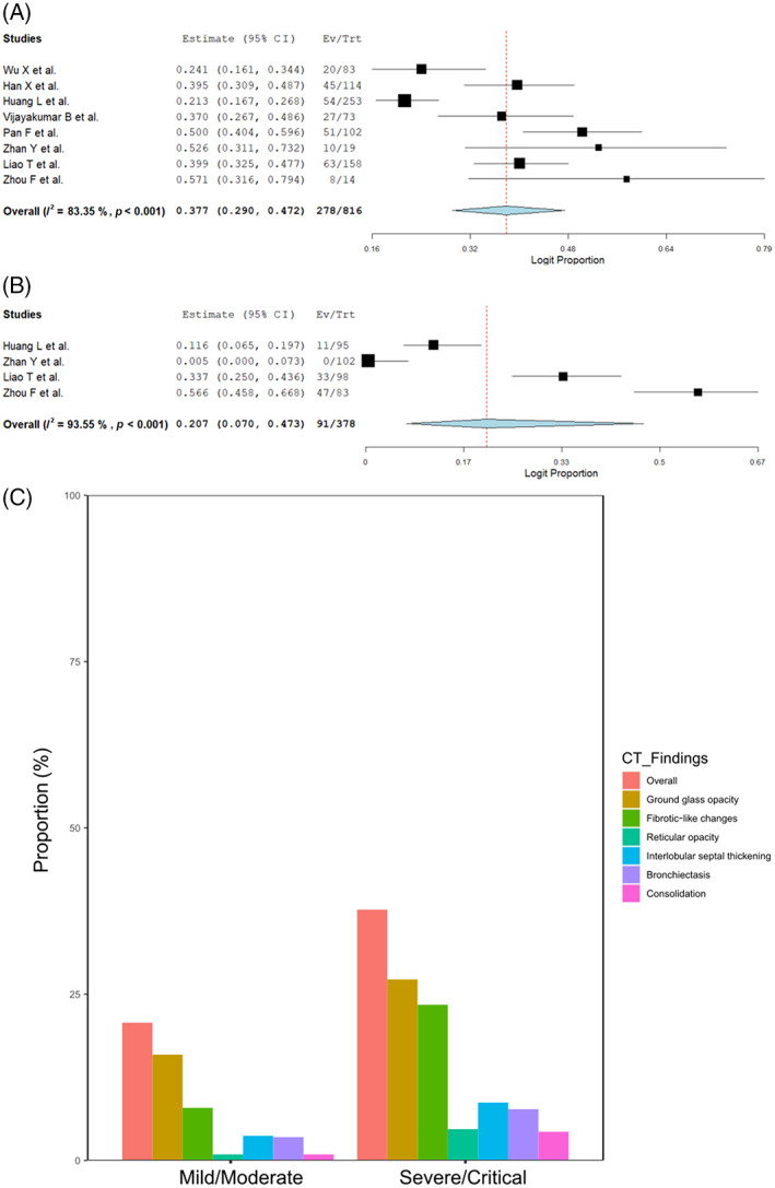
FIGURE 3.

(A) Forest plots of overall residual computed tomography (CT) abnormalities at long‐term follow‐up in severe/critical patients. (B) Forest plots of overall residual CT abnormalities at long‐term follow‐up in mild/moderate patients. (C) Bar graph showing the differences in the proportion of each chest CT finding between severe/critical and mild/moderate patients at long‐term follow‐up
