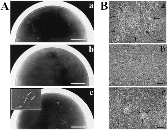
FIG. 1.
Screen for suppressor mutants of galU S. flexneri. Shown are macroscopic (A) and microscopic (B) views of plaque formation by S. flexneri strains 2457T (wild-type) (a), SS100 (ΔgalU2) (b), and SSW301 (ΔgalU2 pepP1) (c) on L2 cell monolayers. Plaques formed by SSW201 (ΔgalU2 cydC1) and SSW401 (ΔgalU2 ribE1) are identical in size and morphology to those formed by SSW301 (data not shown). (A) Plaques were photographed at 72 h postinfection. The inset in panel c is magnified 4×; arrows in panel c indicate plaques. Bar, 1 cm. (B) Plaques were photographed at 48 h postinfection. Arrows indicate the periphery of a single plaque in each panel. Bar, 100 μm.

