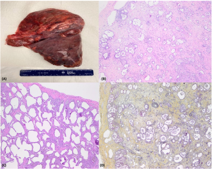
FIGURE 2.

(A) Gross image of the right lung with congestion, subpleural cystic dilatation involving upper lobe, likely secondary to barotrauma. (B) Histopathology (H & E ×10) demonstrating diffuse interstitial expansion with proliferation of fibroblasts, myofibroblasts, and occasional lymphocytes with reactive type 2 pneumocyte hyperplasia a focal non‐specific pattern of lung injury. (C) Diffuse interstitial expansion with lymphocyte predominant inflammatory infiltrate (H&E ×10). (D) Foci of Interstitial expansion with predominance of mature collagenous fibrosis (Movat pentachrome staining)
