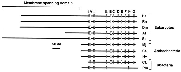
FIG. 4.
Structures and conserved motifs of the HMG-CoA reductase proteins. Location of conserved motifs I to IV and A to G are indicated by shaded and open boxes, respectively. Pm indicates the HMG-CoA reductase from P. mevalonii. Other abbreviations used are as in Fig. 3.

