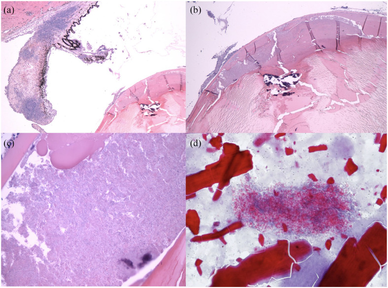
Figure 3.
Histopathology. (a) Case 3, hematoxylin and eosin, × 2 magnification showing lymphoplasmacytic iritis. (b) Case 3, hematoxylin and eosin, × 4 magnification showing regionally severe equatorial lens fiber degeneration. (c) Case 3, hematoxylin and eosin, × 40 magnification showing innumerable Encephalitozoon cuniculi organisms within the lens, with swollen lens fibers/Morgagnian globules (top center) and a few neutrophils outside the capsule (upper left). (d) Case 1, histopathology. Ziehl–Neelsen acid fast stain, × 60 magnification, lens material and E cuniculi organisms. Images (a), (b) and (c) courtesy of Dr Christopher Reilly, DACVP. Image (d) courtesy of Dr Barbara Nell

