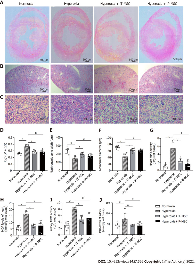
Figure 6.
Beneficial effects of human umbilical cord-derived mesenchymal stem cells administration in the heart and kidneys of hyperoxic neonatal rats. A: Representative images of harvested heart sections stained with HE for morphometric analyses (scale bars = 500 μm); B and C: Representative photomicrographs of the hematoxylin and eosin stained sections of kidneys obtained at 40 × magnification (scale bars = 200 μm) and 200 × magnification (scale bars = 100 μm) respectively; D: Fulton’s index (right ventricle/left ventricle + interventricular septum) was measured to quantify the degree right ventricular hypertrophy (n = 5); E and F: Nephrogenesis was assessed through measuring the width of the nephrogenic zone and the glomerular diameter (n = 7); G–J: Myeloperoxidase and malondialdehyde levels were measured to evaluate the degree of inflammatory and oxidative reaction in heart and kidney tissues respectively (n = 7). aP < 0.05; bP < 0.01; cP < 0.001. iT: Intratracheal; iP: Intraperitoneal; RV: Right ventricle; LV: Left ventricle; IVS: Interventricular septum; MPO: Myeloperoxidase; MDA: Malondialdehyde; MSC: Mesenchymal stem cell.

