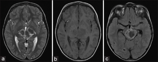
Figure 1.

Axial T2 (a), T1 (b) weighted MRI images of the brain show hypointense area involving bilateral thalami with a hyperintense rim on T2 weighted images. Axial FLAIR image (c) shows a hypointense area in the midbrain with a hyperintense rim. Few other similar characteristic lesions are seen in the right temporal and occipital lobe
