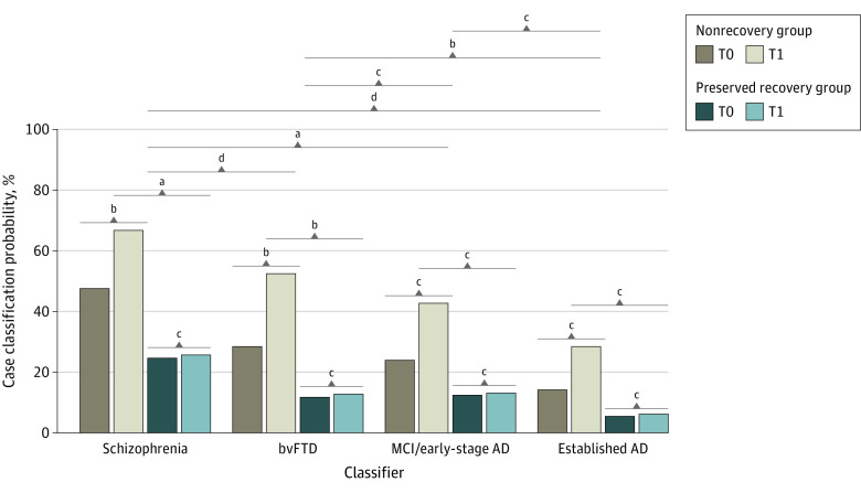
Figure 4. PRONIA Longitudinal Magnetic Resonance Imaging Analysis Describing the Development of Case Classification Likelihoods Between the Baseline and Follow-up Magnetic Resonance Imaging Data of the PRONIA Nonrecovery and Recovery Samples.
Likelihood changes over time were compared by means of generalized estimating equations including Brain Age Gap Estimation as a covariate. Results of estimated marginal means analyses conducted for the functional trajectory, time point, and classifier factors were visualized. See eTable 11 in Supplement 1 for a tabular representation of results. AD indicates Alzheimer disease; bvFTD, behavioral-variant frontotemporal dementia; MCI, mild cognitive impairment; T0, baseline visit; T1, 1-year follow-up visit.
aP < .01.
bP < .05.
cNot significant.
dP < .001.

