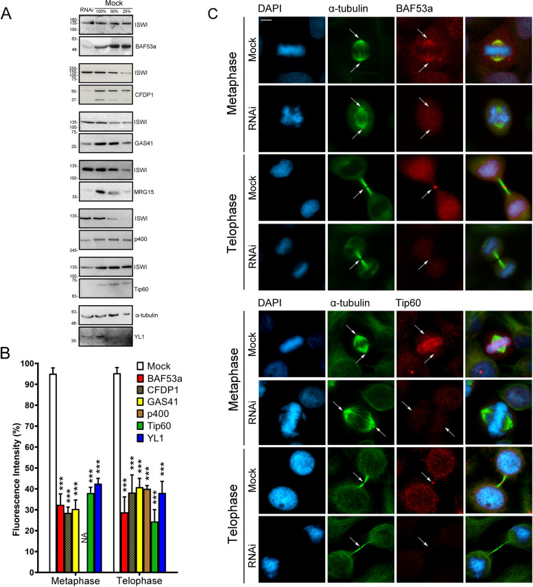
Fig. 3.
Validation of antibodies against the CRS. A Total CRS amount assessed by Western blotting in RNAi-treated and mock-treated cells. B Histograms showing the fluorescence intensity of immunostaining in RNAi-treated and mock-treated cells. The colors of bars are referred to Fig. 1B. Bars with different dashed lines indicate SRCAP complex-specific subunits (CFDP1) or p400/Tip60-specific subunits (p400 and Tip60). Plain colored bars indicate subunits common to both complexes (BAF53a, GAS41, and YL1). The fluorescence intensity showed a decrease ranging from about 60 up to 75% in RNAi-treated HeLa cells compared to the mock-treated cells. Fluorescence intensity was assessed using the ImageJ software and statistical significance was verified by T- test. C Examples of IFM staining. The fluorescence intensity of BAF53a and Tip60 clearly decreased in RNAi-treated compared to mock-treated cells. Scale bar = 10 μm

