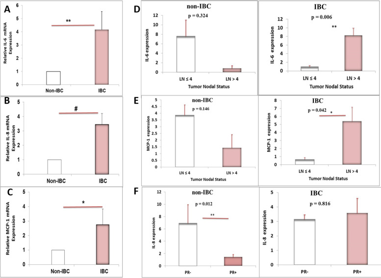
Fig. 3.
qRT-PCR comparing the expression level of the predominant cytokines secreted by CAAT isolated from non-IBC versus IBC patients. A A significantly higher mRNA expression level of IL-6, (B) IL-8, and (C) MCP-1 of CAAT ex-vivo culture from patients with IBC vs. non-IBC by 4.17, 3.46, and 2.75 folds, respectively. ** P < 0.01, # P < 0.0001.* P < 0.05 as determined by unpaired Student t-test. Quantification of target genes mRNA was expressed relative to the housekeeping gene (GAPDH) mRNA. All bar graphs represent fold change+/−SEM (n of non-IBC = 21 and n of IBC = 11). D A significant up-regulation in IL-6 mRNA expression of obese IBC patients that had lymph node (LN) metastasis (n = 10, P = 0.006) in the right panel and no change in IL-6 mRNA expression of obese non-IBC patients that have LN metastasis (n = 17, P = 0.324) in the left panel was observed. E The transcript level of MCP-1 is significantly up-regulated in obese IBC patients that have LN metastasis (n = 10, P = 0.042) in the right panel, but not in obese non-IBC patients that have LN metastasis (n = 17, P = 0.146) in the left panel. F A significant down-regulation in IL-8 mRNA expression of obese non-IBC patients that express PR receptor (n = 21, P = 0.012) in the left panel but not in obese IBC patients that express PR receptor (n = 10, P = 0.816) in the right panel

