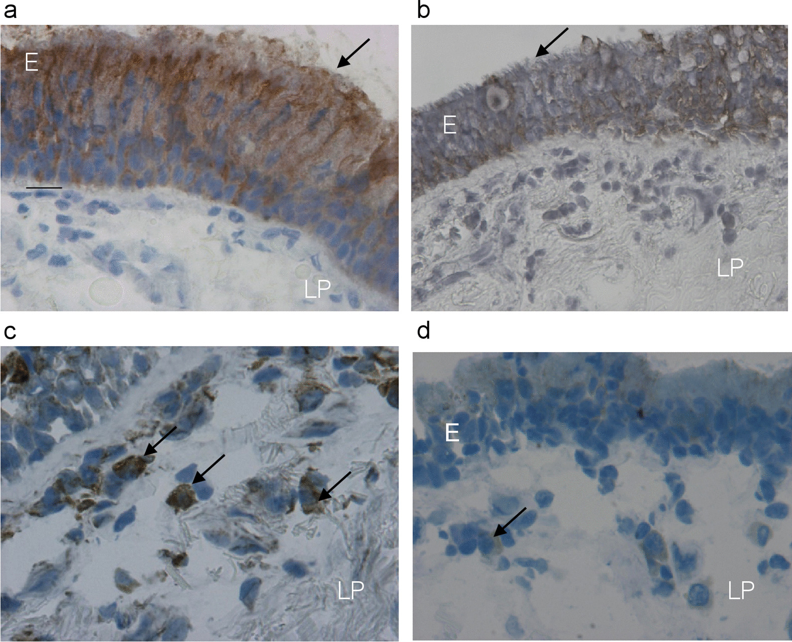
Fig. 2.

Photomicrographs showing bronchial mucosa from a representative non-decliner (a, c) and a rapid decliner (b, d) with COPD, immunostained for identification of secretory IgA protein (a, b) and plasma cells (c, d) in the epithelium (E) and bronchial lamina propria (LP). Results are representative of 21 non-decliners and 15 rapid decliners. Arrows (a, b) indicate immunopositivity in epithelial cells, which was reduced in rapid decliners (b), and immunostaining of plasma cells (c, d), which were reduced (d) in the lamina propria of rapid decliners with COPD. Bar = 20 micron
