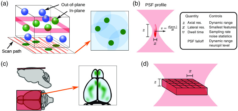
Fig. 1.
Optical imaging basics. (a) Laser scanning microscopy (e.g., two-photon microscopy) focuses the laser on a point inside the tissue, sequentially illuminating a chosen plane. Neurons intersecting the plane show up in the rendered images, whereas out-of-plane neurons accumulate as background, and combined nonsomatic activity (e.g., smaller dendritic and axonal components) as neuropil. (b) Choices in the optical illumination impact image properties. The image resolution is defined by the PSF, which is determined by microscope optics and optical properties of the sample. The dwell time controls the time spent at each location and is related to the scanning speed, i.e., the framerate, and can influence the number of photons collected per pixel and thus the signal strength. In laser scanning microscopy, these, in combination with scanning and laser technology, the image size, resolution, framerate, and noise characteristics of the image are determined. (c) Widefield imaging simultaneously illuminates the entire cortical surface plane, capturing dynamics across multiple brain areas. (d) In widefield imaging, camera characteristics primarily determine the image acquisition size and rate. The optical resolution for widefield imaging is strongly limited by microscope optics and optical aberrations from the sample.

