Fig. 4.
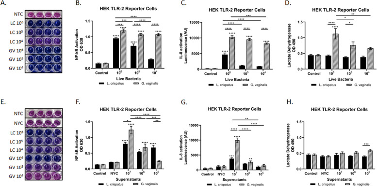
L. crispatus and G. vaginalis activate NF-κB signaling through TLR2; however, only G. vaginalis results in increased IL-8 levels. The HEK TLR2 reporter cell line was used to determine if either live or bacteria-free supernatants from L. crispatus and G. vaginalis activated TLR2-mediated cell signaling. Representative images (A, E) and the corresponding quantification (B, F) of the QUANTI-Blue NF-κB detection assay, IL-8 activation (SEAP quantification) (C, G), and cytotoxicity (D, H) were all altered after exposure to live bacteria and bacteria-free supernatants from L. crispatus and G. vaginalis. For A and E, darker blue/purple indicates higher NF-kB. Values are mean ± SEM. Asterisks over the individual bars represent comparisons with control; asterisks over solid lines represent comparisons between treatment groups. *p < 0.05, **p < 0.01, ***p < 0.001, ****p < 0.0001

