Fig. 7.
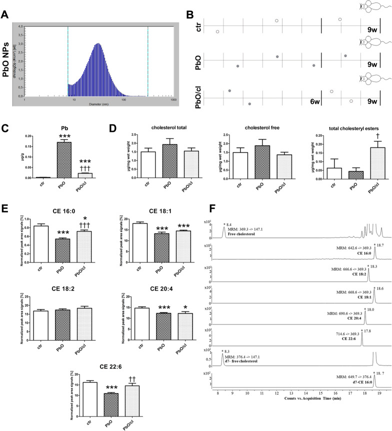
Cholesterol and cholesteryl esters in liver after 9-week PbO NP inhalation. A Particle number-size distribution of PbO NPs in the inhalation chambers measured by Scanning Mobility Particle Sizer (SMPS) in the 2nd experiment. B Design of the inhalation experiment. One group of animals inhaled clean air (ctr) for a period up to 9 weeks, the second group inhaled air with PbO NPs (PbO), and the third group inhaled air with PbO NPs for 6 weeks and thereafter clean air for following 3 weeks (PbO/cl—clearance group). Symbols of light circle indicate clean air, and symbols of dark circles indicate PbO NPs. C Analysis of Pb concentration (µg/g) in blood. Limit of detection in the blood was 0.003 µg/g Pb. The graphs values indicate average ± SD for 5 mice/group; ***p < 0.001 compared with the corresponding control group (ctr), and †††p < 0.001 compared with the corresponding PbO NP group by unpaired t-test. D Quantification of total cholesterol, free cholesterol and total CEs analyzed in liver samples measured by Cholesterol/Cholesteryl Ester Quantitation Kit. The values in graphs indicate average ± SD for 4–5 mice/group; †p < 0.05 compared with the corresponding PbO NP group by unpaired t-test. The amount of cholesterols given in µg was normalized to 1 mg of liver (wet weight). E LC–MS quantification of selected cholesteryl esters (CEs) analyzed in liver samples of control mice (ctr), mice inhaled PbO NPs (PbO) and clearance group of mice (PbO/cl). The graphs values indicate average ± SD for 4–5 mice/group; *p < 0.05; ***p < 0.001 compared with the corresponding control group (ctr), and ††p < 0.01, and †††p < 0.001 compared with the corresponding PbO NP group by unpaired t-test. The absolute abundance of free cholesterol is given in µg/mg wet weight. The value is normalized to signals of deutered internal standard and 1 mg of liver (wet weight). The relative quantitative responses of individual CEs are given as a peak area signal normalized to signal of internal standard and 1 mg of wet weight of liver (expressed as a percentage). F Representative extracted ion chromatograms (EIC-MRM) of LC-ESI MS/MS separation of selected lipids and internal deuterated standards used in all experiments. Several abundant species of CEs were extracted from livers of control mice. LC–MS data were obtained by using optimized experimental conditions as described in the "Methods" section. Retention times and typical MRM transitions used for quantification are shown for each compound

