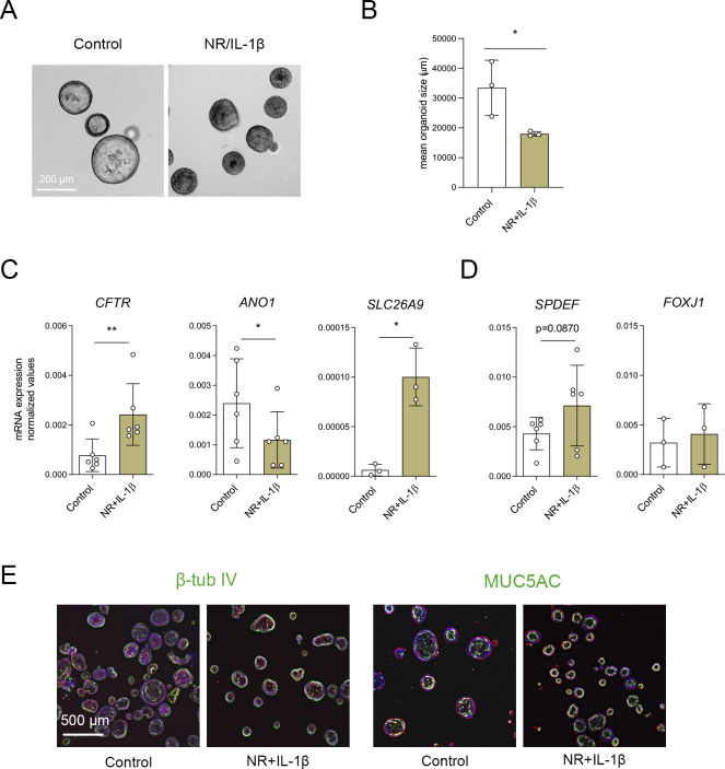
Figure S4. Characterization of nasal-airway organoids (NAOs) cultured with NR/IL-1β.
(A) Brightfield images of cystic fibrosis (CF) F508del/F508del NAOs cultured in control conditions or with NR+IL-1β for 5 d. (B) Quantification of the mean organoid area (μm2, means ± SD) of control and NR+IL-1β cultured CF F508del/F508del NAOs (n = 3 independent donors). (C, D) mRNA expression analysis of CF F508del/F508del NAOs (n = 3–6 independent) that were left unstimulated (control) or cultured with NR+IL-1β for 5 d. (C, D) mRNA expression was determined of (C) CFTR, ANO1, SLC26A9 (all chloride channels), (D) SPDEF (secretory cells), and FOXJ1 (ciliated cells). Results represent target mRNA expression normalized for the geometric mean expression of the housekeeping genes ATP5B and RPL13A (means ± SD). (E) Immunofluorescence staining of CF organoids (control and cultured with NR+IL-1β for 5 d) was conducted of β-tubulin IV (ciliated cell) and MUC5AC (goblet cell) (green). DAPI (blue) was used for nuclear staining. Phalloidin (red) was used for actin cytoskeleton staining. (B, C, D) Analysis of differences was determined with a paired t test (B, C, D). *P < 0.05, **P < 0.01.

