Fig. 1.
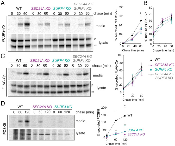
PCSK9 secretion requires SEC24A and SURF4. (A) PCSK9-V5 maturation and secretion were examined in WT and KO cell lines by pulse–chase with [35S]methionine. PCSK9-V5 was immunoprecipitated with α-V5 from lysates and conditioned media at the indicated times and detected by SDS-PAGE and autoradiography. Percentage of secreted PCSK9 was calculated as [band intensity in media at a given time point]/[total protein intensity (media + lysate) at time 0]. PCSK9 is detected as proprotein (p) and mature (m) bands. Plots show the mean ± SD of three independent experiments. Statistical test was a one-way ANOVA with Dunnett’s correction for multiple comparisons, *P < 0.05. (B) Percentage of mature PCSK9 was calculated as [band intensity of the lower MW (mature) species at a given time point]/[total protein (mature + proprotein + secreted) at time 0]. Plots show the mean ± SD of three independent experiments. Statistical test was a one-way ANOVA with Dunnett’s correction for multiple comparisons; no significant difference was detected. (C) FLAG-Cp secretion was examined in WT and KO cell lines. FLAG-Cp was immunoprecipitated with α-FLAG from lysates and conditioned media at the indicated times and detected by SDS-PAGE and autoradiography. Autoradiographs are representative of three independent experiments. As in A, secretion of FLAG-Cp was quantified by phosphorimage analysis. FLAG-Cp is detected as precursor (p) and mature (m) bands. Plots of the mean ± SD are shown in the right-hand panel. (D) Endogenous PCSK9 secretion from the indicated HuH7 cell lines was quantified by pulse–chase with [35S]methionine. PCSK9 was immunoprecipitated with α-PCSK9 from lysates and conditioned media at the indicated times and detected by SDS-PAGE and autoradiography. Plots of the mean ± SD of three independent experiments are shown in the right-hand panel, *P < 0.05. PAGE, polyacrylamide gel electrophoresis.

