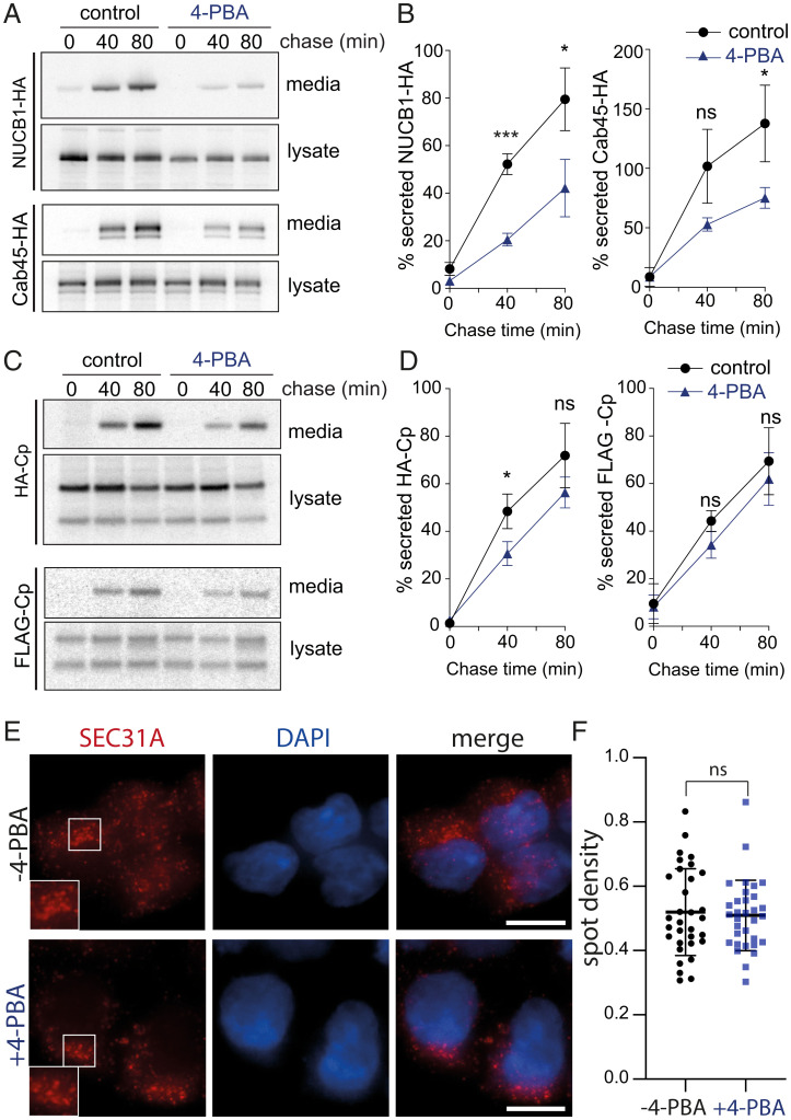
Fig. 6.
4-PBA selectively inhibits secretion of SURF4 clients but not a bona fide bulk flow marker. (A and C) Secretion of the indicated proteins was examined in the presence of 10 mM 4-PBA, which was added during the starvation phase of a pulse–chase experiment and maintained over the course of the experiment. NUCB1-HA, Cab45-HA, and HA-Cp were immunoprecipitated with α-HA from lysates and conditioned media at the indicated times and detected by SDS-PAGE and autoradiography. For FLAG-Cp, α-FLAG was used for immunoprecipitation. (B and D) Percentage secretion into the media was quantified by phosphorimage analysis. Plots show the mean ± SD of three independent experiments. Statistical test was an unpaired t test, *P < 0.05, ***P < 0.001. (E) HEK-293T cells were left untreated or incubated with 10 mM 4-PBA for 4 h and then fixed, immunostained for Sec31A, stained with DAPI, and subjected to fluorescence microscopy. Insets in the left panels show higher magnification of the boxed areas. Scale bars, 10 µm. (F) Quantification of the density of SEC31A-postivie puncta in the presence and absence of 4-PBA. Values represent the number of SEC31A-positive puncta identified per cell. A total of 32 cells from each condition were measured. Error bars represent SD; statistical test was an unpaired t test. ns, not significant; PAGE, polyacrylamide gel electrophoresis.

