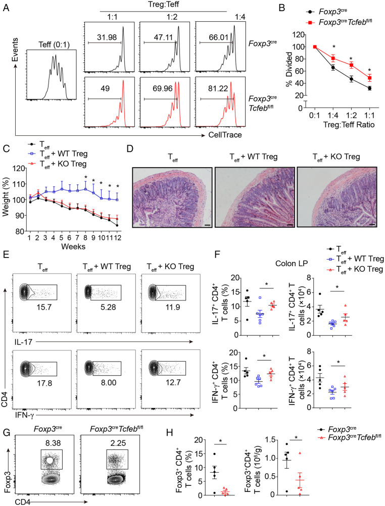
Fig. 3.
TFEB is critical for Treg suppressive function. (A and B) Cell trace-labeled WT naïve CD4+ T cells were stimulated with anti-CD3/anti-CD28 (1 µg/mL of each) in the absence or presence of Treg cells sorted from Foxp3YFP-Cre mice or Foxp3YFP-CreTcfebfl/fl mice at indicated ratios for 72 h. Representative histograms (A) and the mean percentages of divided cells (B) were shown. (C–H) Rag2−/− mice received 4 × 10⁵ CD4+CD25−CD45RBhigh T cells alone or in combination with 2 × 10⁵ WT or Tcfeb-knockout Treg cells. (C) Weight changes of mice post T cell transfer were shown (n = 5 or 6). (D) Representative images of colon sections. (Scale bars: 100 µm.) (E, F) Representative flow cytometry plots and frequencies of IL-17- and IFN-γ-producing CD4+ cells in colonic LP. (G, H) Representative flow cytometry plots and frequencies of CD4+Foxp3+ Treg cells in the spleens. *P < 0.05. Data are representative of two independent experiments with similar results. Data are means ± SEM and were analyzed by two-tailed, unpaired Student’s t test.

