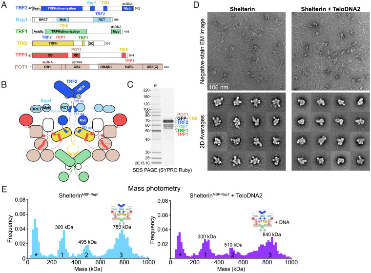
Fig. 4.
Structural heterogeneity within shelterin. (A) Domain schematics for TRF2, Rap1, TRF1, TIN2, TPP1, and POT1 with amino acids indicated. BRCT, BRCA1 C-terminal domain; RCT, Rap1 C-terminal domain. (B) Cartoon schematic for dimeric shelterin. Coloring is as in Fig. 1A. (C) SDS-PAGE of purified reconstituted shelterin. Protein gel as in Fig. 1B. (D) Negative-stain EM analysis of shelterin purified in the presence or absence of ds–ss junction telomeric DNA (TeloDNA2). EM images displaying raw particles are shown above reference-free 2D class averages. Class averages showing a high signal-to-noise ratio were selected for display. (E) Mass photometry of reconstituted shelterin containing MBP-Rap1 and GFP-TIN2 after an amylose pull down. Data were taken in the presence or absence of exogenously added TeloDNA2 as indicated. Peaks are numbered and approximate maxima are indicated. Asterisks indicate a peak arising from minor buffer contaminants.

