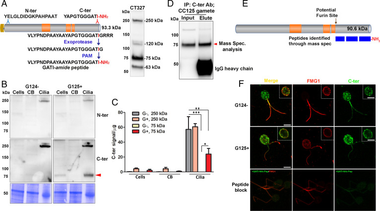
Fig. 1.
Processing and localization of proGATI in minus and plus gametes. (A) Diagram shows Cre03.g204500 (preproGATI) with Prorich regions in orange. The N-terminal and C-terminal peptides used as antigens are shown above the diagram. The pathway leading to C-terminal amidation is illustrated below the diagram. The immunoblot to the right of the diagram shows the bands detected by serum from one of the rabbits (CT327) inoculated with a mixture of N-ter and C-ter peptide conjugates (SI Appendix, Fig. S1). (B) Immunoblot of cells, deciliated cell bodies (CB) and cilia of minus (G124−) and plus (G125+) resting gametes using affinity-purified proGATI N-ter and C-ter antibodies. Equal amounts of protein (20 µg) were loaded. (C) Quantification of C-ter signal revealed significant enrichment of both 250-kDa and 75-kDa bands in cilia. Results are the average of duplicates. Means were compared with ± range. Asterisks indicate significant differences between groups: *P < 0.05, **P < 0.01, ***P < 0.001. (D) Affinity-purified C-ter antibody was used to immunoprecipitate cross-reactive material from mating type plus (CC125) gamete cell lysates. The 75-kDa fragment (red arrowhead) was excised and analyzed by mass spectrometry. (E) Only peptides between Met697 and Ile904-amide were identified, covering 76.4% of this region; all of the spectra identified from the C terminus were amidated. (F) Maximal projection confocal images of minus and plus resting gametes stained with the C-ter proGATI antibody (green) and antibody to FMG1 (red). Insets show a single Z-plane. Plus gametes probed with antibody preincubated with the GATI-amide peptide (Peptide block) exhibit reduced staining (green) in cell bodies and cilia (Bottom). Similar localization of proGATI in gametes was obtained in three independent experiments. (Scale bars, 5 µm apply to all panels.)

