Fig. 2.
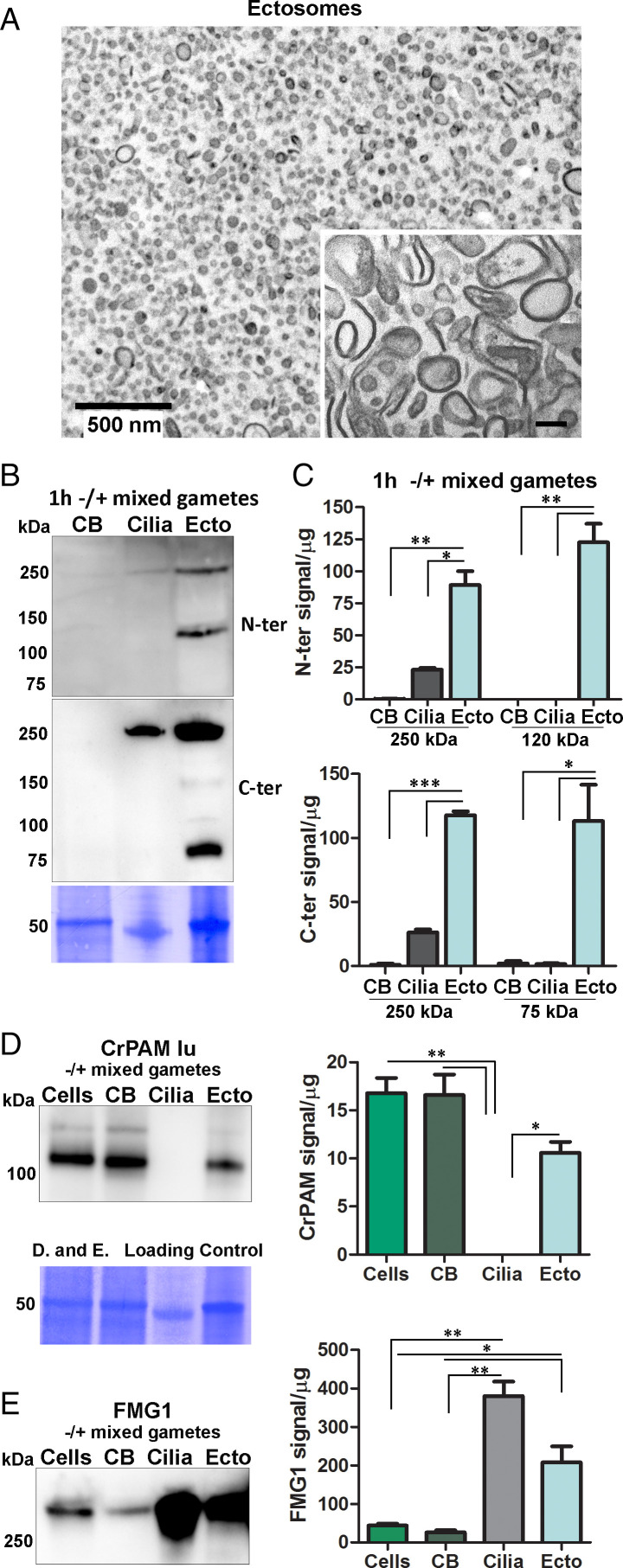
Processing of proGATI in mating ectosomes. (A) Cross-section transmission electron micrograph of an agarose-embedded ectosome pellet isolated from 1 h -/+ mixed gametes. Inset shows a higher magnification image of ectosomes treated with Na2CO3 to remove peripheral membrane proteins. (Scale bar, 100 nm.) Images are representative of three independent experiments. (B) Deciliated cell bodies (CB), cilia, and ectosomes (Ecto) isolated from mixed gametes were fractionated by SDS/PAGE, blotted, and probed with the affinity purified N-ter and C-ter proGATI antibodies. (C) Graphs showing enrichment of N-ter and C-ter signals for the 250-kDa band and smaller fragments. Results are average of two independent experiments; mean ± range. Asterisks indicate a statistically significant difference between two groups: *P < 0.05, **P < 0.001, ***P < 0.0001. Immunoblots from the same membrane showing CrPAM (D) and FMG1 (E) levels in cells, cell bodies (CB), cilia and ectosomes (Ecto) isolated from mixed gametes; quantification was as in C. The loading control applies to both panels D and E.

