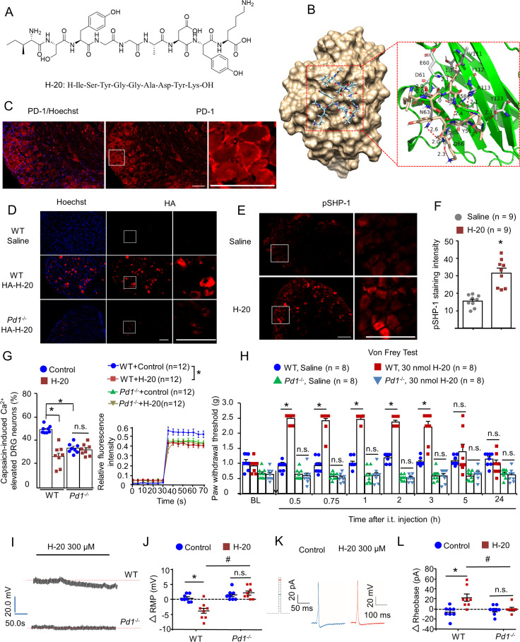
Fig. 1.
A peptide ligand of PD-1. (A) Chemical structure of H-20. (B) Binding mode of H-20 with PD-1 (PDB ID: 4ZQK). (C) Expression of PD-1 on DRGs. (Scale, 50 μm.) (D) Intrathecal HA–H-20 (30 nmol, 1 h), the in vivo binding of HA–H-20 with PD-1 on DRGs. (Scale, 50 μm.) Four mice per group. (E and F) Intrathecal H-20 (30 nmol, 1 h) increased SHP-1 phosphorylation in naïve WT mice. (E) pSHP-1 immunostaining in DRGs. (Scale bar, 50 µm.) (F) Immunofluorescence intensity of pSHP-1 in DRGs. Four mice per group, nine images per group. *P < 0.05, versus saline group. Student’s test. (G) Pretreatment with H-20 (300 μM) attenuates capsaicin-induced increase in [Ca2+]i in cultured DRGs via PD-1. (Left) The proportion of neurons responsive to capsaicin. Mean ± SEM *P < 0.05, versus WT control group. Eight independent experiments for per group. Two-way ANOVA. (Right) The typical traces of calcium responses. Twelve neurons per group. *P < 0.05, versus WT control group. Two separate three-way repeated measures (RMs) ANOVA (0 to 30 and 35 to 70 min). (H) H-20 modulates basal mechanical pain thresholds via PD-1. Mean ± SEM *P < 0.05, versus saline group. Eight mice per group. Three-way RM ANOVA. (I–L) H-20 modulates neuronal excitability via PD-1. Mean ± SEM eight neurons per group. *P < 0.05, versus WT + control group. #P < 0.05, versus WT + H-20 group. n.s., no significance. Two-way ANOVA. (I and J) H-20 alters hyperpolarization of the RMP in WT mice, but not in Pd1−/− mice. (K and L) H-20 increases the rheobase in cultured DRGs in WT mice, but not in Pd1−/− mice.

