
FIGURE 3.
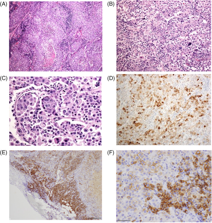
(A–C) Hematoxylin and eosin staining showed that tumor cells included atypical large cells and prominent nucleoli. Tumor cells also exhibited neuroendocrine architectural features, such as palisading features and necrotic areas (A, magnification ×40; B, magnification ×200; C, magnification ×400). (D–F) Immunostaining showed that tumor cells were diffusely positive for (D) chromogranin (magnification ×200), (E) CD138 (magnification ×100), and (F) CD138 (magnification ×400)