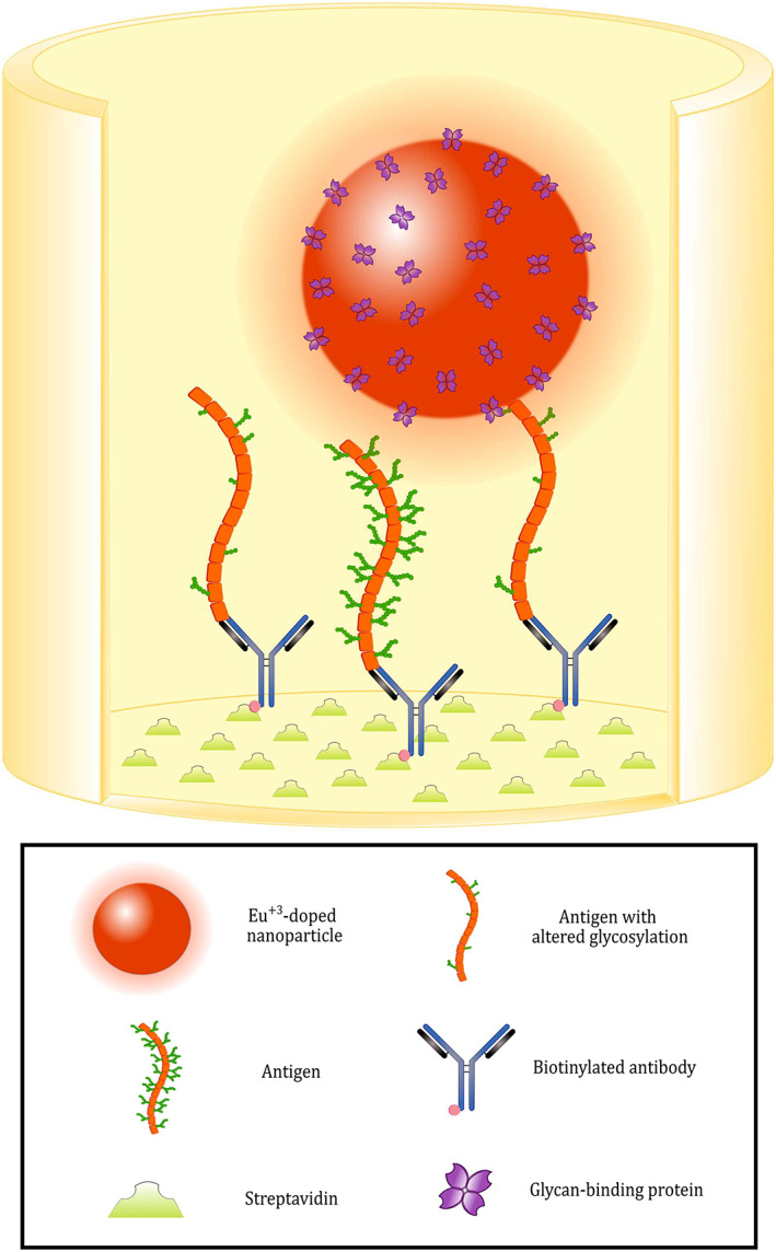
FIGURE 1.

Schematic representation of the glycovariant assay principle. The biotinylated capture antibodies are immobilized on the surface of streptavidin‐coated yellow microtiter wells. The target antigen is then recognized by the capture antibody. In the final step, Eu+3‐doped nanoparticles coated with glycan‐binding proteins (lectins or antibodies) are added to the wells: the proteins coated on their surface recognize altered glycans on the cancer‐related target antigen. Between each step, the wells are washed to remove the unbound fraction of assay components. The measurement of time‐resolved fluorescence is performed on a spot on the bottom of each well
