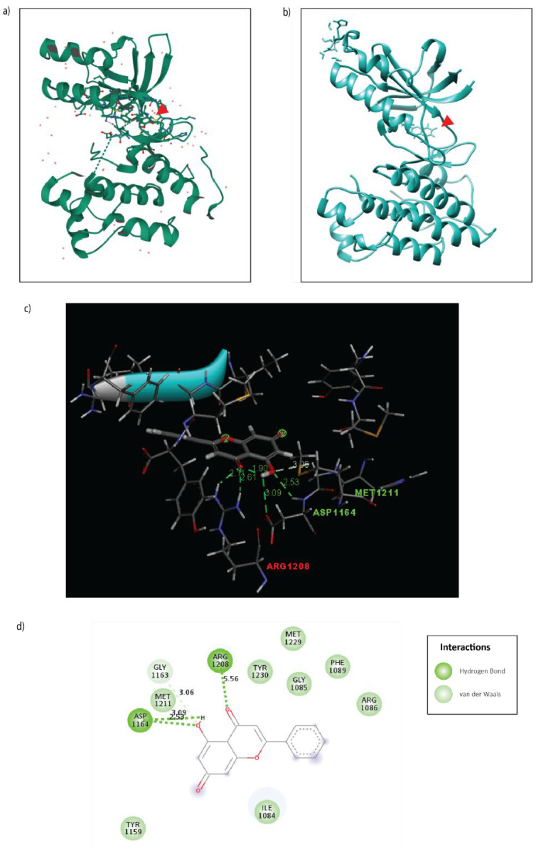
Figure 5.
Molecular docking of Galangin to the tyrosine kinase domain of the c-Met receptor. (a) 3D X-ray crystallography structure of c-Met inhibitor, AM7, bound to c-Met (PDB: 2RFN). (b) 3D Docking model of Galangin (PDB: 57D docked to the c-Met receptor (PDB:1R1W) using SwissDock. (c) 3D interaction plot of Galangin binding to c-Met receptor with distance in (Å). (d) 2D interaction plot of Galangin binding to c-Met receptor. The important interactions were highlighted, including conventional hydrogen bond (dark green dot line) and van der Waals (light green).

