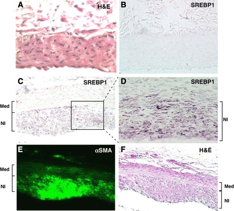
FIGURE 3.
SREBP-1 is increased in the neointima following stent placement. A stent was deployed in rat abdominal aortas for 4 weeks. Subsequently, vessels were harvested and analyzed by immunohistochemistry and immunofluorescence. Six μm sections were stained as described below. A, Control normal rat abdominal aorta stained with H&E reveals no neointima but a medial and adventitial (top) layer. B, Immunohistochemical staining for SREBP-1 in normal control untreated rat abdominal aorta is undetectable. C, Following stent placement for 4 weeks, a neointima layer is now visible and robust staining of SREBP-1 is detected. D, Higher magnification of SREBP-1 staining within the neointima. E, Immunofluorescence of FITC-anti-αSMA reveals robust staining within the neointima. F, H&E staining of the abdominal aorta following 4 weeks of stent placement. Reprinted with permission from Zhou et al.22 Copyright American College of Cardiology Foundation. All permission requests for this image should be made to the copyright holder. FITC indicates fluorescein isothiocyanate, H&E, hematoxylin and eosin; NI, neointima; SREBP1, sterol regulatory element-binding protein 1; αSMA, α-smooth muscle actin.

