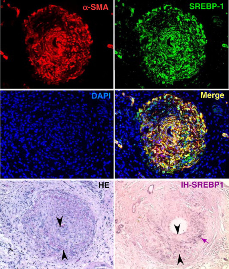
FIGURE 4.
SREBP-1 is increased in both the neointima and VSMC layer of mouse carotid arteries following vascular injury using a model of angioplasty. Four weeks after probe injury, carotid arteries were harvested and subjected to immunofluorescence or immunohistochemistry as indicated. Sections of 6 μm were stained with anti-SREBP-1 or Cy3-anti-α-SMA or stained using H&E. Nuclei were stained with DAPI in blue. Merge image of SREBP-1 (green) and α-SMA (red) is shown in yellow. SREBP-1 detected using immunohistochemistry is represented in purple and indicated by the small arrow. Neointima is indicated as the area between arrowheads in the H&E-stained and immunohistochemistry sections. In uninjured arteries from the same animals, SREBP-1 was barely detected under the same conditions by both methods (data not shown). Reprinted with permission from Zhou et al.22 Copyright American College of Cardiology Foundation. All permission requests for this image should be made to the copyright holder. DAPI indicates 4',6-diamidino-2-phenylindole; H&E, hematoxylin and eosin; IH-SREBP-1, immunohistochemistry for SREBP-1; SREBP1, sterol regulatory element-binding protein 1; VSMC, vascular smooth muscle cell; αSMA, α-smooth muscle actin.

