
Fig. 9.
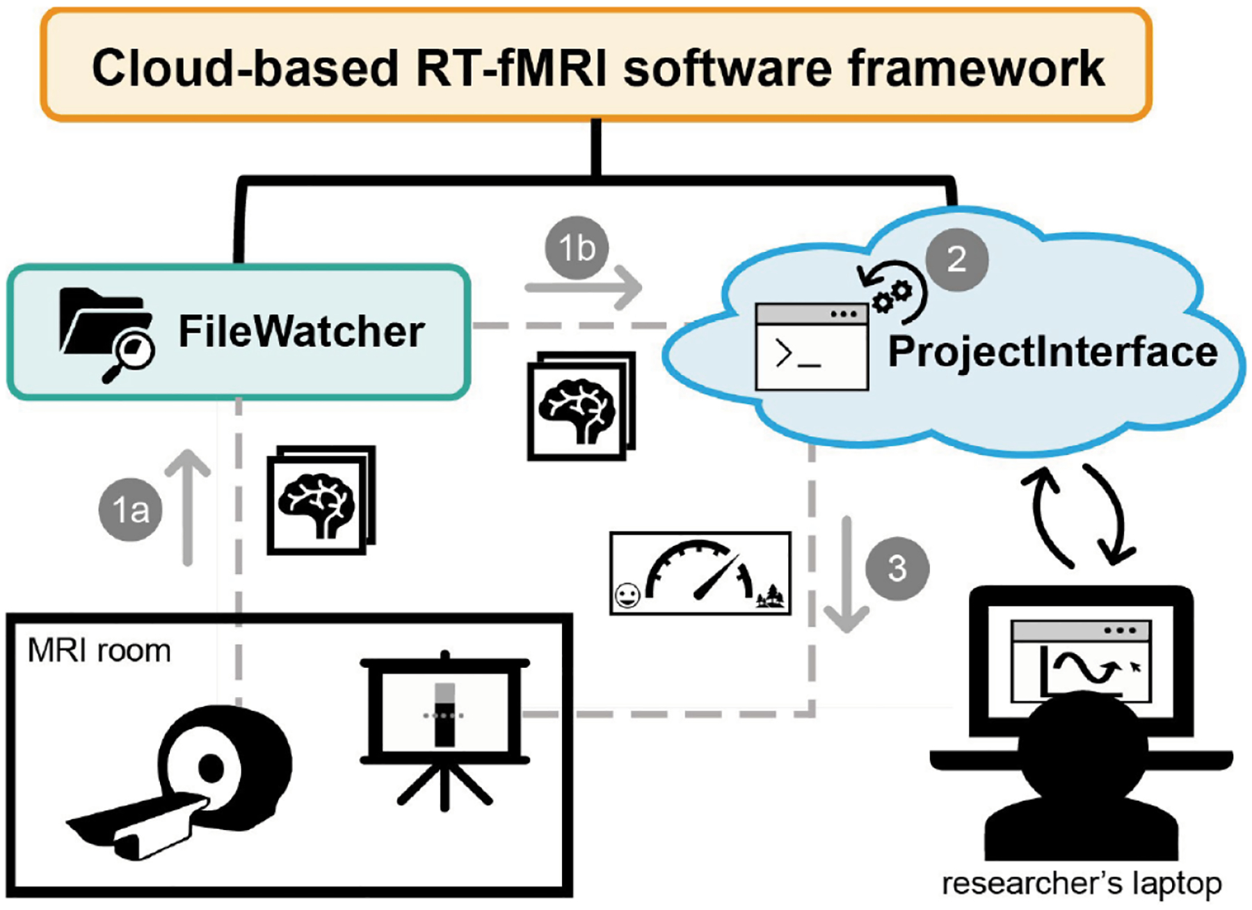
Schematic of our cloud-based software framework for real-time fMRI experiments. The framework has two main components: the FileWatcher and the ProjectInterface. (1a) The FileWatcher watches for the arrival of new DICOM images on the scanner computer and (1b) forwards the image to the ProjectInterface, running on the cloud. (2) The ProjectInterface, which wraps the experimenter’s code, processes the DICOM data and runs the experimenter’s analysis code to obtain a measure of the participant’s brain state. The experimenter accesses the cloud application from a browser page that can run on a laptop. Among other things, the experimenter can initiate/finalize the session, change settings, and even observe the graph output of the analysis results. (3) The analysis results are provided to the participant as visual neurofeedback presented on the projector in the MRI room.