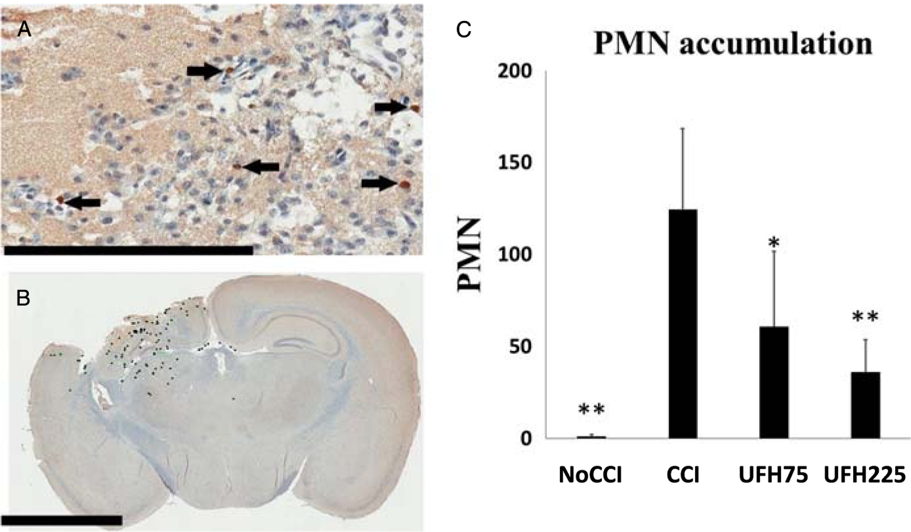
Figure 4.

(A) Representative immunohistochemistry images of coronal brain sections demonstrating PMNs in the contusional/pericontusional regions as 1A8-positive cells indicated by black arrows (scale bar 200 μm). (B) Mean PMN counts were quantified by a blinded observer using annotated whole-brain sections in the coronal plane at the level of the contusion (scale bar 3 mm). (C) Compared with untreated injured animals, both UFH treatment groups demonstrated significantly less accumulation of PMNs in penumbral brain tissue. *p < 0.05, **p < 0.01 versus CCI.
