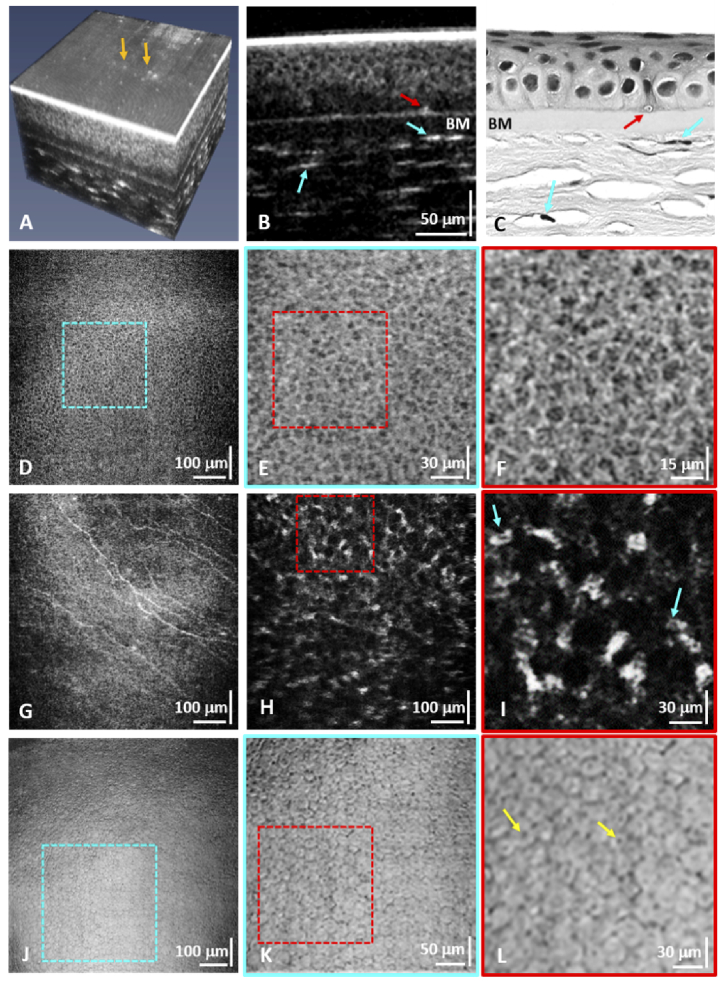
Fig. 3.
(A) Flattened 3D LS-SD-OCT image of the anterior cornea from a healthy volunteer. (B) Cross-sectional image of the anterior cornea showing the cellular structure of the epithelium, the Bowman's membrane, cross-sections of sub-basal corneal nerves (red arrow), and keratocyte nuclei in the anterior stroma (blue arrows). The same morphological features are resolved in the cross-sectional H&E corneal histology ex-vivo (C). (D) Enface image of the corneal epithelium with magnified views (E, F) of the regions of interest (ROIs) marked with the blue and red dashed lines that show individual epithelial cells. (G) Enface images of sub-basal corneal nerves. (H, I) Enface images of the anterior stroma showing keratocyte nuclei. (J) Enface maximum intensity projection image of the corneal endothelium. (K, L) Zoomed ROIs showing individual endothelial cells.

