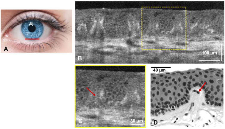
Fig. 4.
(A) Human healthy inferior limbus (imaged location marked with red line). (B) Cross-sectional LS-SD-OCT image that shows the POVs and the cellular structure of the limbal crypts located in between the POVs. (C) Magnified view of the ROI marked with yellow line in (B). (D) H&E histological image of the a single palisade and the surrounding limbal crypts.

