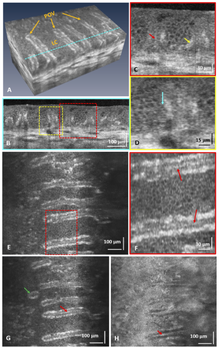
Fig. 5.
(A) Flattened volumetric image of the healthy inferior human limbus showing the palisades of Vogt (POVs) and the cellular structure of the limbal crypts (LC). (B) Cross-sectional image of the POVs and the LC corresponding to the location in (A) marked with the blue dashed line. (C, D) Zoomed images of the ROIs marked in (B) that show pigmented cells along the inner borders of the limbal crypts (yellow arrow), hyper-reflective structure under the POVs (red arrow), and small sized cells, probably limbal stem cells or transient amplifying cells, located along the crypt walls (blue arrow). (E, G, and H) Enface images of the POVs and LC acquired at different depth locations. (F) Zoomed view of the ROI marked in (E). These images show a focal stromal projection (green arrow) and hyper-reflective line structures, possibly blood vessels or nerves, located under the POVs (red arrows).

