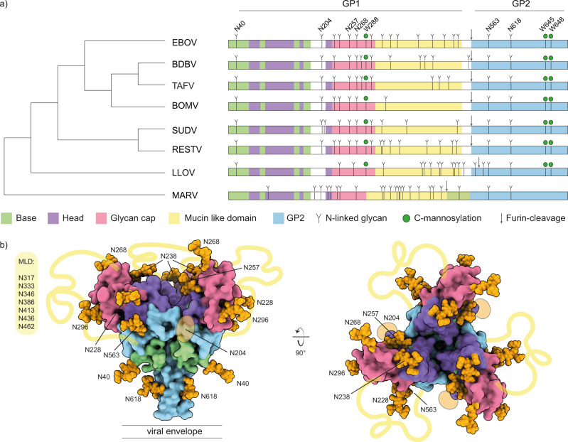
Fig. 1. Sequence and structure analysis of Filovirus GP glycosylation.
a Schematic of filovirus GP domain structure with annotated N-linked glycosylation and C-mannosylation. The cladogram on the left is based on the full GP sequences. Domain coloring as indicated below the diagram. Conserved N-linked glycans in ebolavirus species are annotated on top. b Pseudomodel of EBOV GP with core pentasaccharide of N-linked glycans shown as orange spheres. Model is built with GLYCAM based on PDB ID 5JQ3. The yellow lines indicate the approximate location of the MLD, connecting the respective termini of the glycan cap and GP2 subunits.

