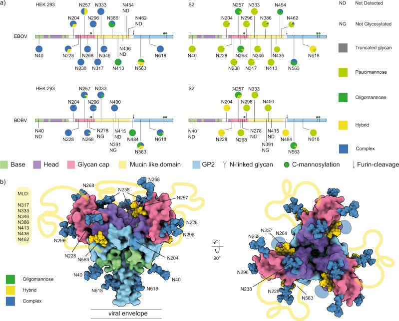
Fig. 2. N-linked glycosylation profiling of ebolavirus GP.
a Overview of site-specific N-linked glycan processing in ebolavirus GPΔTM from HEK293 and S2 cells as determined by LC-MS/MS. The glycans were classified by HexNAc content as truncated, paucimannose, oligomannose, hybrid or complex. Shown is the average of a duplicate experiment. b Pseudomodel of EBOV GP (as in Fig. 1), with glycans colored by main class (oligomannose, hybrid, complex) as observed in HEK293 cells.

