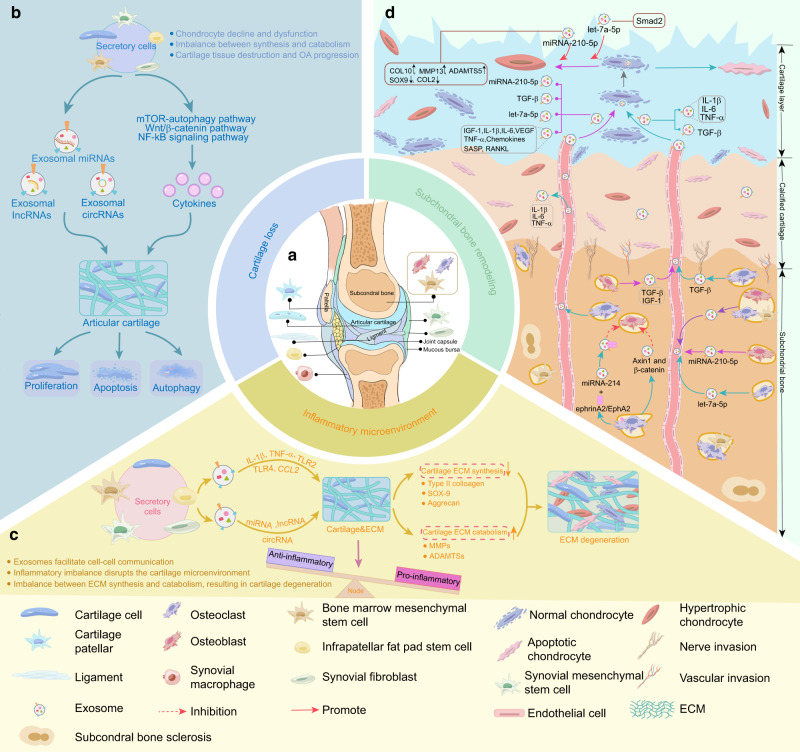
Fig. 4.
The major biological functions of different tissue-derived exosomes in OA. a Exosomes can be released by various periarticular cells, including chondrocytes, osteoblasts, osteoclasts, and bone marrow MSCs in subchondral bone, synovial fibroblasts, macrophages, and MSCs in the synovium and infrapatellar fat pad. b Exosomes can mediate cell-to-cell communication to regulate multiple phenotypes of chondrocytes, including proliferation, apoptosis, and autophagy, thus affecting cartilage damage. c Inflammatory factors, noncoding RNAs, and other mediators released by exosomes from different cells contribute to the remodeling of the inflammatory microenvironment and drive ECM catabolism. d Exosomes and cytokines produced by osteoblasts, osteoclasts, and bone marrow MSCs in subchondral bone are transported to the cartilage layer and regulate chondrocyte metabolism. In addition, exosomes secreted by osteocytes can participate in subchondral bone remodeling, leading to changes in the biomechanical properties of OA subchondral bone, ultimately causing damage to the cartilage. OA osteoarthritis, MSCs mesenchymal stem cells, IL-1beta interleukin-1β, IL-6 interleukin-6, TNF-α tumor necrosis factor-alpha, CCL2 chemokine (C-C motif) ligand 2, TLR2 Toll-like receptor 2, TLR4 Toll-like receptor 4, MMP13 matrix metalloproteinase 13, ADAMTS5 a disintegrin and metalloproteinase with thrombospondin motifs 5, ECM extracellular matrix, MMP matrix metalloproteinase 13, SOX9 SRY-box transcription factor 9, mTOR mechanistic target of rapamycin, Wnt/β-catenin Wnt-beta-catenin, NF-κB nuclear factor kappa-B, VEGF vascular endothelial growth factor, TGF-β1 transforming growth factor-β1, IGF-1 insulin-like growth factor-1, COL2 type 2 collagen, COL10 type 10 collagen, RANKL receptor activator of NF-κB-associated phenotype β1

