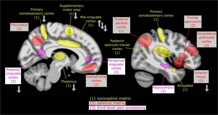
FIGURE 2.
Basic view of ascending pain processing, delineated on two brain sections, with emphasis on early, sub-conscious (1, “nociceptive matrix”: the posterior operculo-insular cortex, the primary sensory areas, p-mid-cingulate cortex, supplementary motor area and the amygdala) and conscious pain perception (2, “salience matrix”: the anterior cingulate cortex, the anterior insula, posterior parietal, prefrontal and orbitofrontal cortices), and (3, “areas of third-order brain activation”: the hippocampus and the anterior and posterior cingulate). Nociceptive cortical processing is initiated in parallel in sensory, motor and limbic areas. Some activation may last longer than voluntary motor reaction. Based on brain response dynamics and models described in Bushnell et al. (42), Garcia-Larrea and Bastuji (70). The alterations in pain-induced responses in the brain of individuals with ASD are shown with gray arrows according to Failla et al. (62), Chien et al. (71), and Gu et al. (72).

