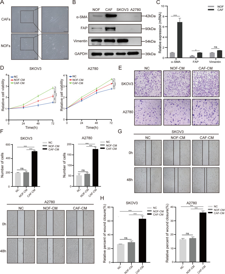
Fig. 1. The conditioned medium (CM) of CAFs promotes proliferation, migration, and invasion of OCCs.
A The typical morphological images of primary CAFs and normal ovarian fibroblasts (NOFs). Bar = 100 μm. B, C Western blotting and RT-qPCR analysis presented the expression of their representative markers (α-SMA, FAP, and vimentin). D CCK8 assay was used to analyze the viability of SKOV3 and A2780 cells co-cultured with CAF-CM and NOF-CM. E, F Cell invasion was assessed by Transwell assay in SKOV3 and A2780 cells after 48-h co-cultured with conditioned medium (100×magnification). G, H Cell migration ability was measured by wound healing assay in SKOV3 and A2780 cells (100×magnification). The quantitative analyses were performed using ImageJ software. Results are presented as the mean ± SD of three independent experiments. *P < 0.05, ***P < 0.001, ns not significant.

