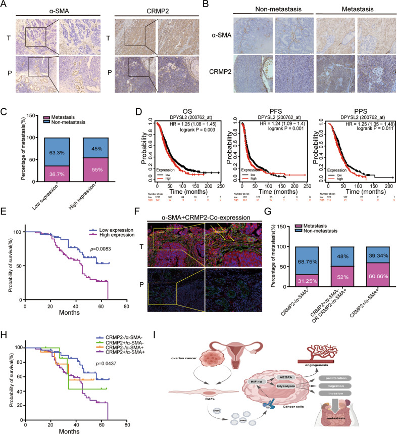
Fig. 6. CRMP2 derived from CAFs contributes to poor prognosis in OvCA patients.
A IHC analysis showed the expression of α-SMA and CRMP2 in epithelial ovarian cancer (EOC) tissues and para-carcinoma tissues (100×magnification, the zoomed-in section, 200×magnification). B Tissue microarray (TMA) containing 118 EOC patients was used to analyze expression of α-SMA and CRMP2 in non-and metastatic cases. C High expression of CRMP2 was correlated with tumor metastases. D, E Both Kaplan–Meier plotter and TMA analyses validated the high expression of CRMP2 that was related to poor prognosis. F There were α-SMA and CRMP2 co-expression in tumor stroma via immunofluorescence analysis (yellow arrow). No signal was detected in para-carcinoma tissues (200×magnification, the zoomed-in section, 400×magnification). Red and green fluorescence signal represented the expression of CRMP2 and α-SMA, respectively. While the yellow fluorescence signal represented co-expression. G, H CRMP2 + /α-SMA + represented the co-expression group, and the double positive group was associated with tumor metastases and probability of survival. I A schematic diagram showed that CRMP2 was secreted from CAFs and then stimulate the down-stream HIF-1α-glycolysis and HIF-1α-VEGFA signaling pathway to regulate the progression of ovarian cancer. OS overall survival, PPS post progression survival, PFS progression free survival.

