Figure 1.
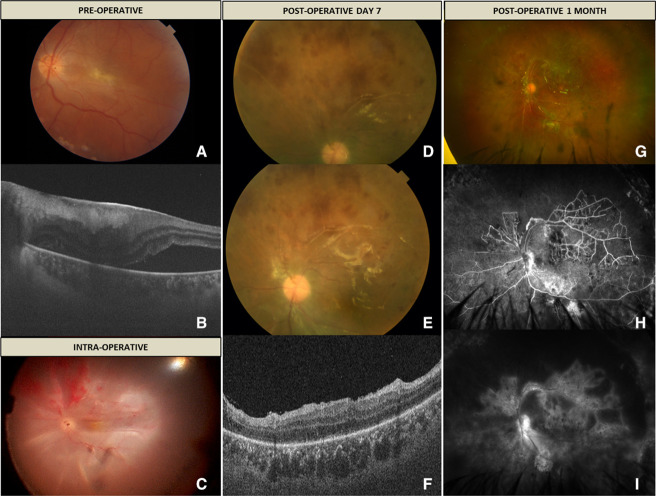
(A) Fundus colour photo of the left eye showing presence of grade 2 epiretinal membrane (ERM) with retinal detachment. (B) Optical coherence tomography (OCT) through the macula shows the presence of ERM with subretinal fluid. (C) Intraoperative photo showing total retinal detachment and presence of occlusive vasculitis and retinal haemorrhages. (D and E) Postoperative colour photo showing signs of active retinal vasculitis in the superior retina. (F) OCT through the fovea of OS showing attached retina and no evidence of ERM. (G) Ultrawide field colour photo showing presence of occluded vessels and resolution of retinal haemorrhages. (H and I) Ultrawide field fundus fluorescein angiography showing extensive peripheral capillary non perfusion (CNP), collateral vessels, and disc and vascular leakage.

