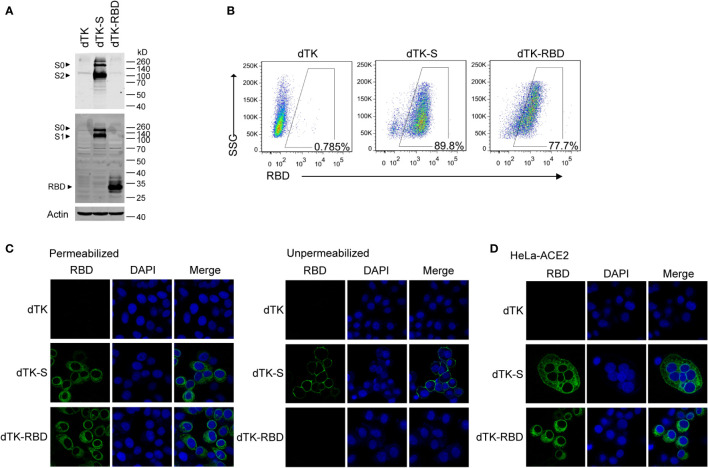
Figure 2.
Expression of RBD and spike protein in cells infected by the recombinant VACV. (A) Vero cells were infected with parent VACV dTK, dTK-S, or dTK-RBD (MOI 0.01) for 24h. Cell lysates were analyzed by Western blotting using S2 or RBD-specific antibodies. (B) Vero cells were infected with VACV dTK, dTK-S, or dTK-RBD (MOI 0.05) for 24h. Cells were permeabilized and stained with anti-RBD antibody, followed by Alexa Fluor 488-conjugated donkey anti-rabbit antibody incubation. Percentages of FITC-positive cells were shown. (C) Immunofluorescence staining of HeLa cells infected with VACV dTK, dTK-S, or dTK-RBD (MOI 0.05) for 24h. Cells were fixed with 4% paraformaldehyde, permeabilized with 0.2% Triton X-100, or stained directly with anti-RBD antibody. Nuclei were stained by DAPI. (D) Immunofluorescence staining of syncytia formed by HeLa cells expressing human ACE2 (HeLa-ACE2). HeLa-ACE2 cells were infected with VACV dTK, dTK-S, or dTK-RBD (MOI 0.05) for 24 h, fixed, permeabilized, and stained with anti-RBD antibody. Nuclei were stained by DAPI. RBD, receptor-binding domain; VACV, vaccinia virus; dTK-S, vaccinia virus-expressing spike protein; dTK-RBD, vaccinia virus-expressing receptor-binding domain; MOI, multiplicity of infection; FITC, fluorescein isothiocyanate.

