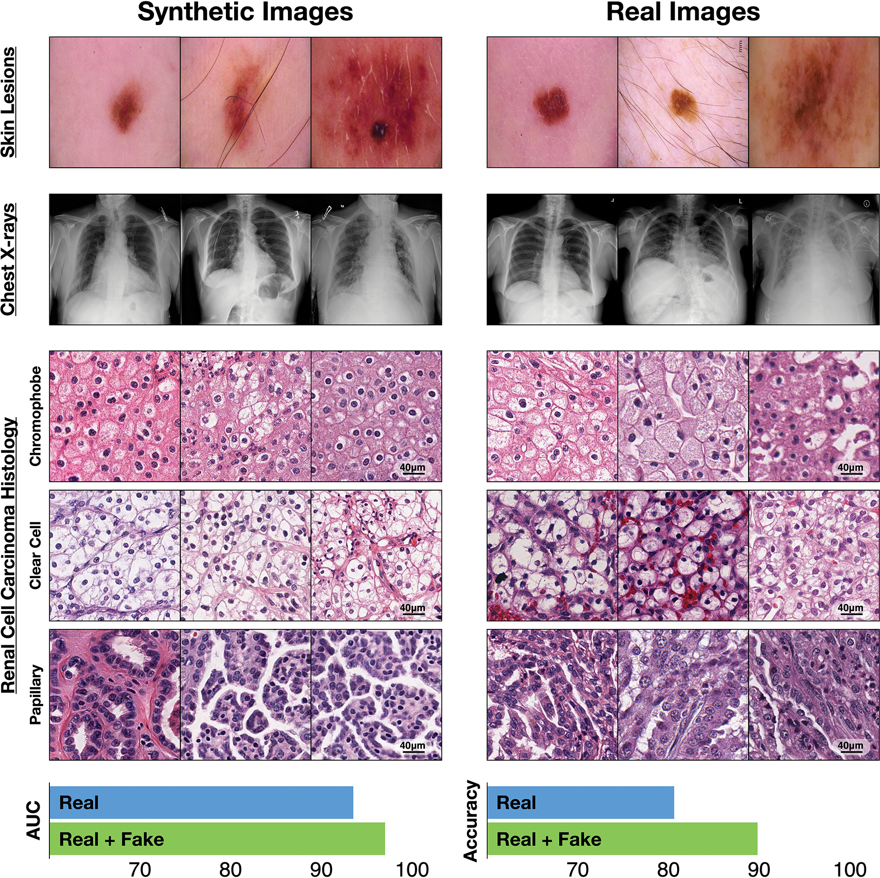
Fig. 1 |. Synthetic medical data in action.

Top, synthetic and real images of skin lesions and of frontal chest X-rays. Middle, Synthetic and real histology images of three subtypes of renal cell carcinoma. Bottom, Areas under the receiver operating characteristic curve (AUC) for the classification performance of an independent dataset of the histology images by a deep-learning model trained with 10,000 real images of each subtype and by the same model trained with the real-image dataset augmented by 10,000 synthetic images of each subtype. Methodology and videos are available as Supplementary Information.
