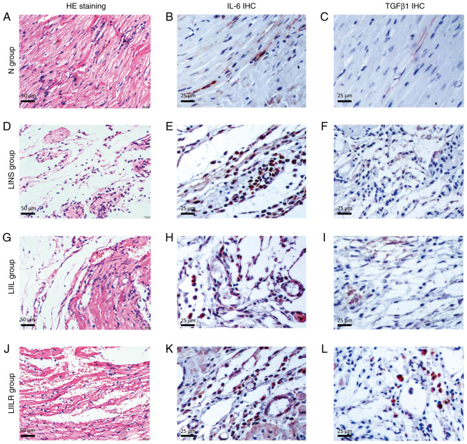
Figure 6.
Histological and immunohistological analyses. (A, D, G and J) HE staining shows the inflammatory response of the cardiomyopathy in rats and myocardial necrosis. The myocardial tissue of (A) the N group showed that the morphology of myocardial cells was complete and the boundaries were clear. The (D) LINS groups and (G) LIIL groups saw a large number of inflammatory cells in particular infiltrate the infarcted area, accompanied by removal of disrupted ventricular tissue. These pathological phenomena were significantly reduced in the (J) LIILR group, and local vascular reactivation and residual heart muscle could be observed. Representative images of immunohistochemical staining of IL-6 (brown blots) in heart tissues of rat at 2 weeks are shown as (B), (E), (H) and (K), respectively. The number of positive expression particles in the LIIL group was the largest, followed by the LINS group and the LIILR, where they were significantly reduced, and the N group, where it was rare. TGFβ1 immunized dyeing in myocardial tissue was sepia particles, mainly distributed in the infarction region, the number of positive expression particles was the largest in the (I) LIIL group, followed by the (F) LINS group, (L) LIILR group significantly reduced and the (C) N group was rare. HE, hematoxylin and eosin; IHC, immunohistochemistry; N group, normal control group; LINS group, LAD ligation with an injection of normal saline group; LIIL group, LAD ligation with an injection of IL-6 recombinant protein group; LIILR group, LAD ligation with injection of IL-6 receptor inhibitor group.

