
Figure 6.
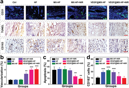
Angiogenesis, apoptosis and inflammation in skin flaps. a) Immunohistochemical staining images of skin flaps with anti‐CD31 antibodies (green) and DAPI (blue), TUNEL staining and CD163 staining in different groups. Scale bars indicate 100 µm (top) and 50 µm (middle, bottom). Quantification of the b) CD31 positive vessels, c) cell apoptosis assessed by TUNEL staining and d) CD163 positive macrophages. n = 6 per group, two‐tailed unpaired Student's t‐tests were performed to calculate the statistical significance between two groups, and *p < 0.05, **p < 0.01, and ***p < 0.001 compared with the control group.