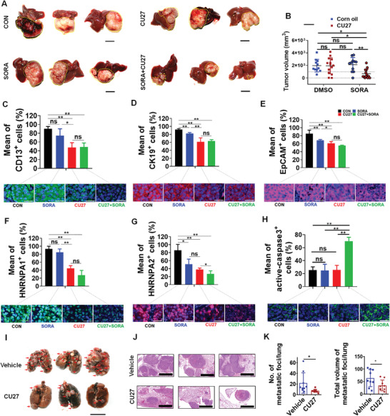
Figure 7.

Therapeutic potential of CU27 in liver orthotopic tumor‐bearing mouse model. A) Representative liver images of intrahepatic xenografted mice in the control group (n = 11), the sorafenib‐monotherapy‐group (n = 11), the CU27‐monotherapy‐group (n = 14), and the combination‐treatment group (n = 14). Bars, 1 cm. B) Statistics of tumor volume between each group. Means ± SD; * p < 0.05, ** p < 0.01; two‐way ANOVA. Representative immunohistochemistry (IHC) staining of C) CD13, D) CK19, E) EpCAM, F) HNRNPA1, G) HNRNPA2 and H) active‐caspase 3 in xenograft tumor tissues (bottom) and the statistics histogram of mean percentage (top). Bars, 30 µm. Means ± SD; * p < 0.05, ** p < 0.01; Student's t‐test. I) Representative lung images of xenografted mice treated with CU27 or vehicle (corn oil). Red arrows, metastatic lung nodules. Bars, 1 cm. J) Representative H&E staining images of metastatic lung foci. Bars, 2000 µm. K) Statistical histogram of the number (left) and total volume (right) of metastatic lung foci. CU27 group (n = 38), vehicle group (n = 37). Six sections per lung were randomly chosen and analyzed in each group. Means ± SD; * p < 0.05; Student's t‐test, excluding the maximum and minimum values for each group.
