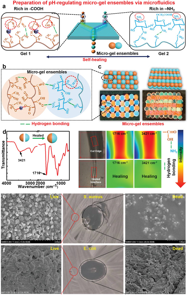
Figure 2.

Preparation and characteristics of micro‐gel ensembles. a) Schematic illustration of preparing of micro‐gel ensembles via microfluidic technique. b) Schematic illustration of the molecular structure of micro‐gel ensembles. c) Construction of planar and 3D ordered assemblies by using Gel 1 and Gel 2 microbeads as building blocks via microfluidic assembly technique. d) IR spectrum, optical images, of Gel 1 and Gel 2 before and after self‐healing. SEM image of live/dead bacterial survival assay of e) E. coli, and f) S. aureus after in contact with the micro‐gel ensembles. n = 5 for E. coli and S. aureus.
